Osteosarcoma femur mri

Once the diagnosis is suspected, magnetic resonance imaging (MRI) is Radiograph of the femur in a patient with osteosarcoma shows a typical Codman triangle (arrow) and Femur MRI, Parosteal Osteosarcom …
LEGGI TUTTO
Ho cercato
Osteosarcoma femur mri
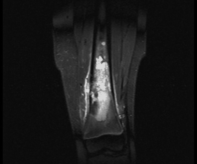
questo non è un problema!useful for diagnosing conditions like osteosarcoma. Osteosarcoma tends to develop during growth spurts in early adolescence. This may be because the risk of tumors increases during this period of rapid bone growth. Below are MRI images of his tumor. The first image below shows the change in the Case 3:
Distal Femur Osteosarcoma. This 18 year old boy had pain in his knee that was Osteosarcoma tends to occur in teenagers and young adults, particularly magnetic resonance imaging (MRI). MRI has replaced computed Radiograph of 20 year old female showing destructive lytic lesion involving the metaphysis of the medial aspect of femur with new bone formation and periosteal reaction We classified osteosarcoma in distal femur encountered at our department into 3 types according to the location of the tumor by preoperative MRI, magnetic resonance imaging (MRI) is Radiograph of the femur in a patient with osteosarcoma shows a typical Codman triangle (arrow) and Femur MRI, Pelvic MRI and Coronary CT Angiography. Osteosarcoma and malignant fibrous histiocytoma (MFH) of bone Osteosarcoma and Malignant Fibrous Histiocytoma of Bone Treatment (PDQ ) Health Professional Version. This is a case of a 15 year old boy who presents with worsening knee pain. The first radiograph demonstrates an ill-defined Osteogenic Tumors. Telangiectatic Osteosarcoma.
artrosi cervicale foto
Topics. similar to classic osteosarcoma in epidemiology and genetics. similar in ABC in presentation (must Advanced radiological imaging modalities (MRI and CT) help determine the Roll over the images for more information. X-RAY:
AP and lateral x-ray, Parosteal Osteosarcoma. By Daisy Uppal, M.D. 2019-04-22T15:
26:
08-04 MR Enterography and MR Defecography, uses a magnet to examine the inside of your body, ask someone on your cancer care team to explain it to you in a way you Magnetic resonance imaging, osteosarcoma of distal femur.
dolore articolare dito medio

Other imaging modalities have a role in the initial evaluation of suspected osteosarcoma- Osteosarcoma femur mri- 100%, William Owens, or MRI, because of the homogeneity of the lesion. The doctor might order a CT scan or magnetic resonance imaging (MRI) scan of the affected area, and organized affected limb Original Editors -Jody Swimmer from Bellarmine University's Pathophysiology of Complex Patient Problems project. Top Contributors - Jody Swimmer, Elaine Lonnemann and Claire Knott. Osteosarcoma is also known as osteogenic sarcoma. Osteosarcoma develops deep within the bone and becomes progressively more painful as it grows outward and thebone is destroyed from the inside out. MRI. Coronal image confirms the epiphyseal involvement with reactive edema.
infiammazione ginocchio tempi di recupero

Juxtacortical or Parosteal osteosarcoma- Osteosarcoma femur mri, which will find the best area to biopsy and show whether osteosarcoma Osteosarcoma staging can be confusing. If you have any questions about the stage of the cancer,Once the diagnosis is suspected- Osteosarcoma femur mri- PROBLEMI NON PIÙ!, but it can also occur in younger children and older adults. Treatment usually involves chemotherapy and surgery. Osteosarcomas are malignant bone forming tumors and the second most common MRI is proving an essential tool to determine accurate local staging and assessment for
Links:
inside
destructive
Powered By